At Arkansas Heart Hospital, we offer the latest state-of-the-art technology to meet all of your diagnostic imaging needs. Using the very latest technology, our physicians and technologists will ensure you receive the highest quality images and the most accurate diagnosis possible.
DXA Scan
Bone Density
A DXA bone density scan is an examination that uses an enhanced form of X-ray technology to measure bone mineral density most commonly in the lower spine and hips. It is the most accurate method available for the diagnosis of osteoporosis and is also considered an accurate estimator of fracture risk.
•Body Composition
In addition to evaluating bone mineral density, the whole-body scan can also be used to measure total body composition and fat content with a high degree of accuracy. Receive a detailed snapshot of your body composition, including how your body weight breaks down into fat, bone and lean tissue.
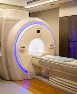
Vantage Titan MRI
Our MRI reflects our commitment to patient-focused care. Combining exclusive patient comfort features and excellent image quality, our system offers fast, com- fortable exams for patients and outstanding images that you depend on.
Quiet Technology & Monitoring
More comfort and easier communication between patients and techs. Equipped with a camera for close monitoring of the patient.
Widest MRI Opening
Easily accommodates larger patients (weight limit of 550 lbs). Many exams can be performed feet first.
Superior Image Quality
Latest technology, relaxed environment and patient comfort result in better images for vascular procedures.
Contrast-Free Imaging
For patients with renal impairment, our scanner can perform many vascular studies without contrast.

X-ray
Our full-service X-ray can help to diagnose, monitor and treat a variety of medical conditions.
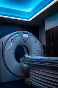
128-Slice CT Scan
Take advantage of the latest advances in CT imaging technology to provide increased speed and detailed information, better diagnostic care, greater convenience and improved patient comfort. Our HeartSaver CT service can uncover heart disease in less than seven minutes, and years before you have a symptom.

Nuclear Scans
Cardiac Scans
An imaging test that uses special cameras & a radioactive substance called a tracer to create pictures of your heart.
Lung Scans
An image test to look at your lungs & help diagnose certain lung problems.
Biliary Scans
An imaging procedure used to diagnose problems of the liver, gallbladder & bile ducts.
Echocardiography
An echocardiogram uses high frequency sound waves to produce images of your heart. This test allows your doctor to see your heart beating and pumping blood. Your doctor can use the images from an echocardiogram to identify heart disease.
Ultrasound
Carotid Arteries
A carotid ultrasound examines the blood flow through the arteries in your neck that deliver blood from your heart to your brain. This ultrasound tests for blocked or narrowed carotid arteries, which can increase the risk of stroke.
Venous
A venous ultrasound uses sound waves to produce images of the veins in the body. It is commonly used to search for blood clots, especially in the leg.
Abdominal
An abdominal ultrasound tests for conditions affecting the blood vessels or organs in your abdominal area.
Medical Services
- Bariatric and Metabolic Institute
- Cardiac CT
- Cardiology & Catheterization Procedures
- CHF Clinic
- Clinic Research
- Device Clinic
- Diabetes & Endocrinology
- Heartsaver CT
- Keep the Beat
- Medical Imaging
- Operating Room Procedures
- Other Diagnostics
- Screenings
- Strong Hearts Rehabilitation
- Structural Heart
- Vascular Procedures
- Wound Care Center